Third Space Endoscopy
and Endoscopic Resection
Peroral Endoscopic Myotomy (POEM)

Submucosal Endoscopy, a New Era of Pure Natural Orifice Translumenal Endoscopic Surgery (NOTES) - Scientific Figure on ResearchGate. Available from: https://www.researchgate.net/figure/Peroral-endoscopic-myotomy-A-Entry-to-submucosal-space-B-Submucosal-tunneling-C_fig5_228083228 [accessed 11 Sep, 2020]
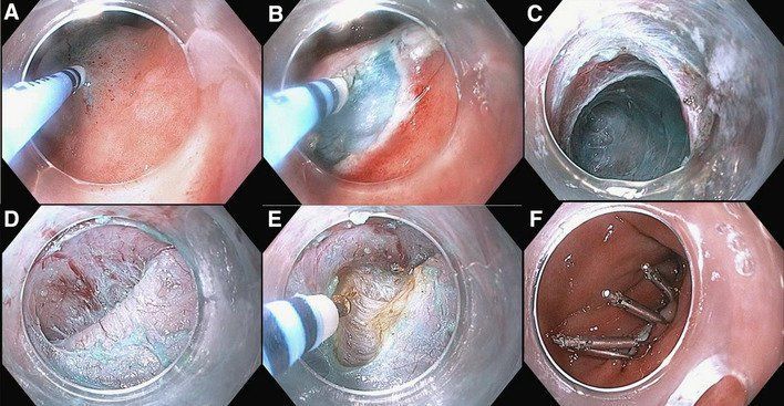
Peroral endoscopic myotomy. (A) Entry to submucosal space. (B) Submucosal tunneling. (C) Endoscopic myotomy, with a total length of 10 cm. (D) Long endoscopic myotomy of inner circular muscle bundles, leaving the outer longitudinal muscle layer intact. (E) Closure of mucosal entry (Adopted from Inoue et al. Endoscopy 2010;42:265-271).16
A Peroral Endoscopic Myotomy (POEM) is performed when a patient is suffering from swallowing disorders such as achalasia. Complications can occur in the esophagus if the muscles contract or tighten abnormally, making it difficult for the patient to swallow food. The procedure is minimally invasive, and is shown to offer faster recovery and less post-procedure pain when compared to its surgical alternative, Heller Myotomy.
The procedure consists of creating a small tunnel within the walls of the esophagus, which will be used by the endoscope to open up and reduce resistance between the muscles of the lower esophageal wall and the gastroesophageal sphincter (the connection point between the stomach and the esophagus). The opening will allow food to pass through the esophagus and into the stomach with ease. After the procedure is completed, the tunnel opening is then closed.
Dr. Michel Kahaleh is a leading physician in the practice of POEM and has performed this operation over 500-times around-the-world. He provided his expertise and has taught the procedure to physicians around Latin America because of the high risk of achalasia in that region.
POEM Procedure as detailed by Dr. Michel Kahaleh.
New Jersey Advanced Endoscopist (2020, March 20) . POEM Procedure - Achalasia treatment [Video]. Youtube. https://www.youtube.com/watch?v=fUoVAGb0AK0&feature=emb_title
Gastric Peroral Endoscopic Myotomy (G-POEM)
Gastric per-oral endoscopic myotomy for refractory gastroparesis: a detailed description of the procedure, our experience, and review of the literature - Scientific Figure on ResearchGate. Available from: https://www.researchgate.net/figure/Steps-of-G-POEM-procedure-A-Selection-of-targeted-gastric-wall-and-creation-of_fig2_323138355 [accessed 11 Sep, 2020]
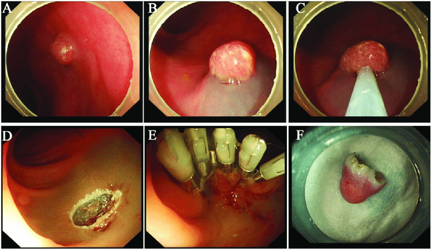
Steps of G-POEM procedure. (A) Selection of targeted gastric wall and creation of submucosal bleb. (B) Longitudinal mucosal incision. (C) Creation of submucosal tunnel. (D) Identifying the PMR. (E) Endoscopic pyloromyotomy. (F) Mucosal access closure using clips.
Gastric Peroral Endoscopic Myotomy (G-POEM) is performed to treat gastroparesis. Gastroparesis occurs when the muscles of the stomach have difficulty contracting, making it difficult for food to leave the stomach and enter the small intestine. The word gastroparesis translates to “a weak stomach,” and the illness can lead to abdominal pain, bloating, nausea, and vomiting.
The procedure is done by creating a tunnel in the stomach wall, using the endoscope to cut the muscles of the pylorus, the connection point between the stomach and small intestine, which will allow food to pass through easier. After the procedure is done, the tunnel opening used by the endoscope is then closed to ensure the integrity of the stomach lumen.
Dr. Michel Kahaleh has co-authored two studies regarding the practice of G-POEM. One of-which has discovered an 85% improvement rate in patients who have undergone the procedure, as well as a decrease in the gastroparesis cardinal symptom index (GCSI) symptom score during follow-ups on such patients.
Zenker’s Diverticulum Per Oral Endoscopic Myotomy (Z-POEM) & Septotomy

Flexible endoscopic and surgical management of Zenker's diverticulum - Scientific Figure on ResearchGate. Available from: https://www.researchgate.net/figure/Images-of-Zenkers-diverticulum-Killian-Jamieson-diverticulum-and-cricopharyngeal-bar_fig1_230755023 [accessed 11 Sep, 2020]
Zenker's diverticulum, Killian-Jamieson diverticulum and cricopharyngeal bar. (A) Zenker's diverticulum, (B) Zenker's diverticulum and Killian-Jamieson diverticulum, and (C) cricopharyngeal bar.
Zenker’s Diverticulum is a condition that affects the patient’s throat, leading to the inability to eat and possible lung infections. Zenker’s Diverticulum is a pharyngeal pouch where a single outpouching of the mucous membrane is located just outside the throat. This pouch will block food from going down into the stomach, and the food collects in the pouch, which can lead to further complications.
Both Z-POEM & Septotomy procedures consist of creating a small incision in the pharyngeal pouch that separates it from the esophageal lumen. Thus, allowing food to pass through the esophagus and prevent the pharyngeal pouch from further accumulating food.
Through Dr. Michel Kahaleh’s many years of performing this operation, he and his fellow physicians at Robert Wood Johnson University Hospital have found overwhelming success. Patients that undergo these procedures enjoy a speedy recovery and return to a normal way of life very quickly.
Endoscopic Mucosal Resection (EMR)
Re-evaluation of indications and outcomes of endoscopic excision procedures for colorectal tumors: A review - Scientific Figure on ResearchGate. Available from: https://www.researchgate.net/figure/The-process-of-endoscopic-mucosal-resection-EMR-A-The-lesion-before-resection-B_fig3_261838912 [accessed 11 Sep, 2020]
The process of endoscopic mucosal resection (EMR). (A) The lesion before resection. (B) Inject saline solution at the submucosa. (C) Release the snare, then re-tighten and resect the lesion. (D) The wound after resection. (E) Seal the wound with metallic clips. (F) The lesion.
Endoscopic Mucosal Resection (EMR) is a procedure that is used specifically to remove lesions located within the Gastrointestinal (GI) tract but does NOT affect areas beyond the outer walls of the GI tract or GI lumen (interior area of a tubular organ which is surrounded by body tissue).
The procedure involved injecting a solution that aims to strengthen and thicken a section of the GI tract, just beneath the location of the lesion. This will create a fluid cushion that allows the lesion to be safely removed. The entire procedure is minimally invasive, making use of an endoscope for its entirety.
This procedure can be safely performed on lesions located within the esophagus, stomach, small intestine, and large intestine.
Endoscopic Full Thickness Resection (EFTR)
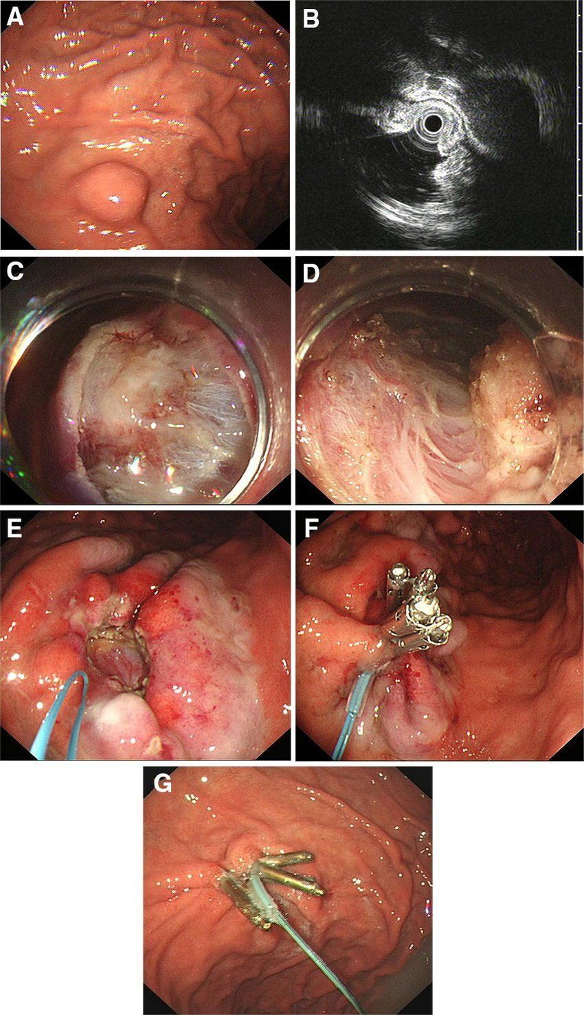
Endoscopic full-thickness resection (EFTR) procedures. (A, B) An oval submocal tumor was located in the middle third of stomach, and EUS [mini-probe] showed it was homogenously hypoechoic orginating from muscularis propria. (C, D) Circumferential incision and deep submucosal dissection. (E, F) After complete removal with full-thickness resecion, the defect was closed with clips and an endoloop. (G) The endoscopic apperarance of the wound at 3 month after EFTR
Endoscopic submucosal excavation and endoscopic full-thickness resection for gastric schwannoma: five-year experience from a large tertiary center in China - Scientific Figure on ResearchGate. Available from: https://www.researchgate.net/figure/Endoscopic-full-thickness-resection-EFTR-procedures-case-2-A-B-An-oval-submocal_fig1_337811329 [accessed 11 Sep, 2020]
Endoscopic Full Thickness Resection (EFTR) is a procedure similar to the above mentioned Endoscopic Submucosal Dissection (ESD). Wherein, a lesion that has affected the GI tract from the lumen through the outer wall is removed from the Gastrointestinal (GI) tract.
This procedure differs from ESD by creating a hole within the GI tract. This hole is then used to remove the lesion. After the procedure, the hole is closed by endoscopic sutures.
This procedure can be safely performed on lesions and tumors located within the esophagus, stomach, small intestine, and large intestine.
Dr. Michel Kahaleh co-authored a study of EFTR on gastric stromal tumors and its effectiveness with just a single procedure.
Endoscopic Submucosal Dissection (ESD)
Endoscopic resection of gastric and esophageal cancer - Scientific Figure on ResearchGate. Available from: https://www.researchgate.net/figure/Endoscopic-submucosal-dissection-ESD-technique-in-early-gastric-cancer-located-at-the_fig1_283325153
[accessed 11 Sep, 2020]
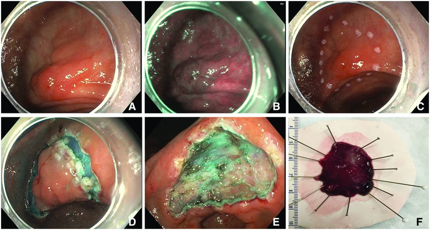
Endoscopic submucosal dissection (ESD) technique in early gastric cancer located at the incisura. (A) Mucosal lesion, spanning approximately 2 cm in white light view. (B) Mucosal lesion, giving cause for concern, in narrow band image view. (C) Perimeter of planned incision marked with electrocautery. (D) After circumferential incision. (E) After completion of dissection. (F) Resection specimen 34 mm x 29 mm.
Endoscopic Submucosal Dissection (ESD) is a procedure that is used to remove tumors or lesions within the Gastrointestinal (GI) tract that have affected the deepest parts of the walls along the GI tract. When compared to Endoscopic Mucosal Resection (EMR), ESD allows the removal of lesions that are much deeper in the GI walls, as well as allowing physicians to remove the targeted lesion in a single piece.
This procedure consists of using a set of special knives to remove the lesion or tumor from the GI walls in a very precise manner. This technique allows the lesion or tumor to be removed with minimal damage to the GI wall, upholding the integrity of the affected organ.
Dr. Michel Kahaleh concludes that this specialty procedure, which was developed in Japan, represents a significant advancement in the field of therapeutic endoscopy. A study co-authored by Dr. Michel Kahaleh documents the success of performing ESD on pediatric patients.
This procedure can be safely performed on lesions and tumors located within the esophagus, stomach, small intestine, and large intestine.
Submucosal Tunneling with Endoscopic Resection (STER)
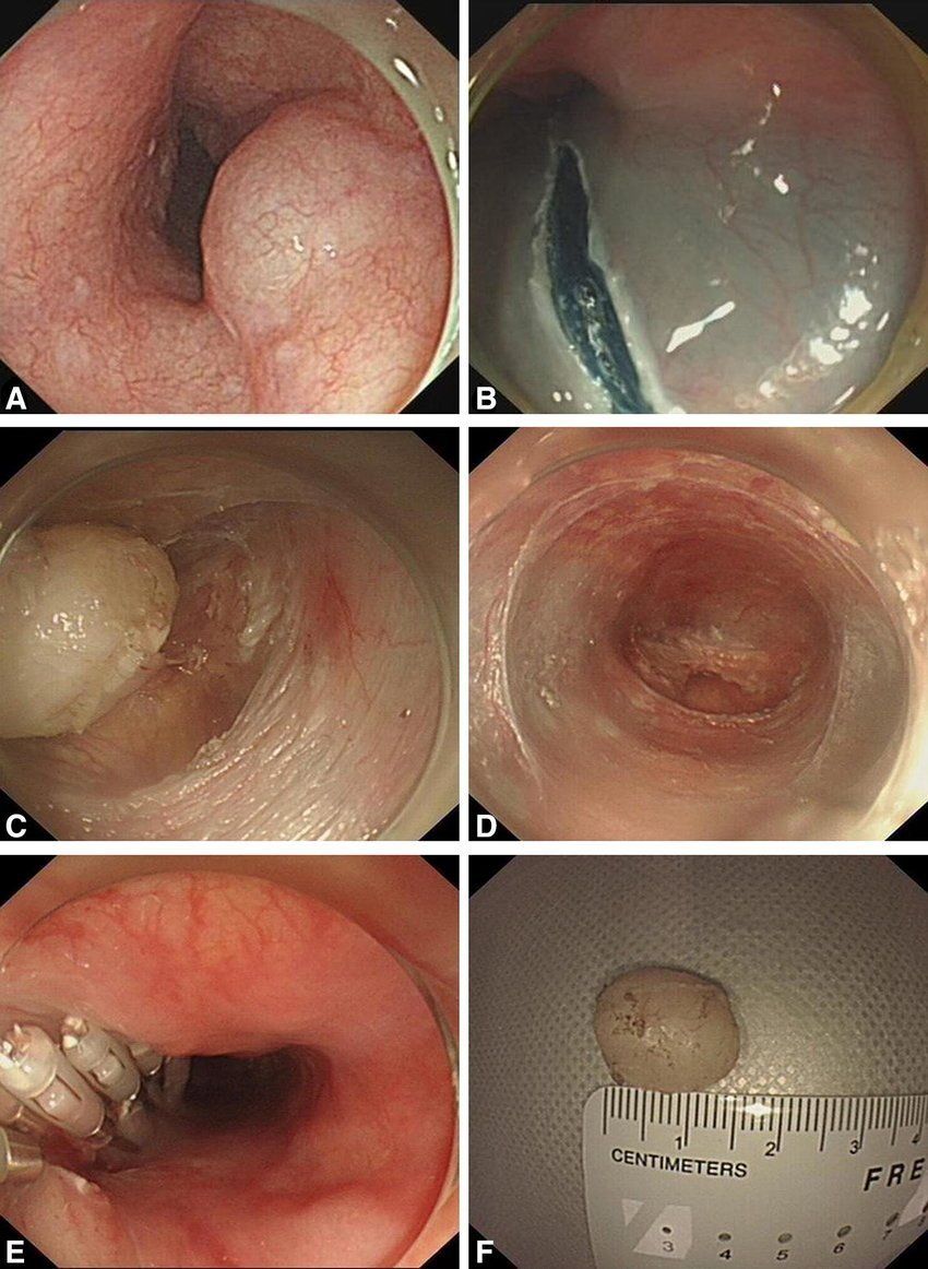
Submucosal tunneling endoscopic resection (STER) to remove an esophageal submucosal tumor (SMT) originating from the muscularis propria (MP) layer. (A) An esophageal SMT was detected by endoscopy. (B) Submucosal injection at 5 cm proximal to the tumor and a 2-cm longitudinal mucosal incision was made as the tunnel entry. (C) Tumor dissection and exposure.
(D) Submucosal tunnel after tumor removal. (E) Closure of the tunnel entry with several clips. (F) Complete resection of the esophageal SMT
The retrospective comparison between submucosal tunneling endoscopic resection and endoscopic submucosal excavation for managing esophageal submucosal tumors originating from the muscularis propria layer - Scientific Figure on ResearchGate. Available from: https://www.researchgate.net/figure/Submucosal-tunneling-endoscopic-resection-STER-to-remove-an-esophageal-submucosal-tumor_fig2_332337858 [accessed 11 Sep, 2020]
Submucosal Tunneling with Endoscopic Resection (STER) is a procedure used to remove tumors from the gastrointestinal (GI) tract. STER, along with Endoscopic Submucosal Dissection (ESD) are the two most widely accepted techniques of removing tumors that have affected the esophagus, stomach, small intestine, and large intestine.
This procedure consists of creating a tunnel within the walls of the GI lumen, which will lead directly to the tumor below. Through the tunnel, an endoscope is sent through and is used to remove the tumor from inside the tunnel. After the procedure, the tunnel is closed off internally by endoscopic sutures.
STER is a minimally invasive procedure that does not require open and shut surgery. Dr. Michel Kahaleh has co-authored two separate studies on Submucosal Tunneling with Endoscopic Resection and is an expert on the procedure.